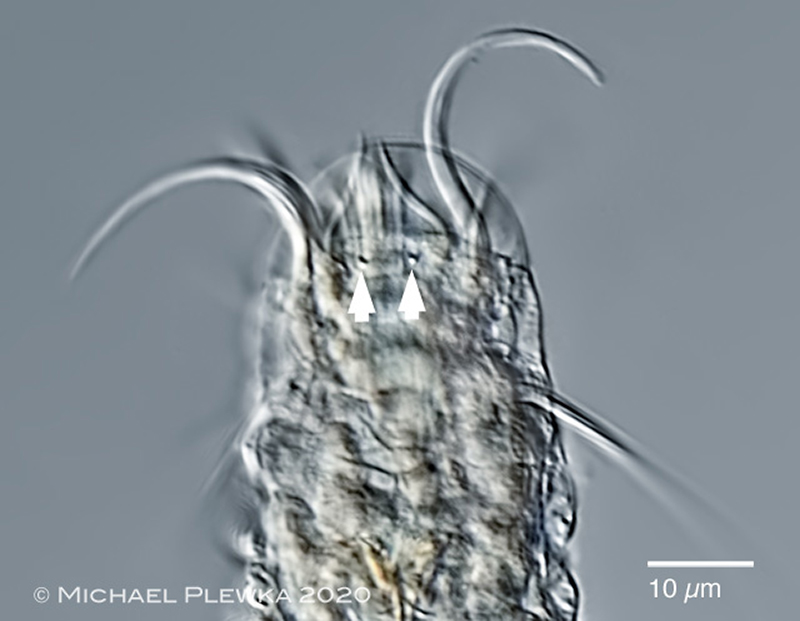
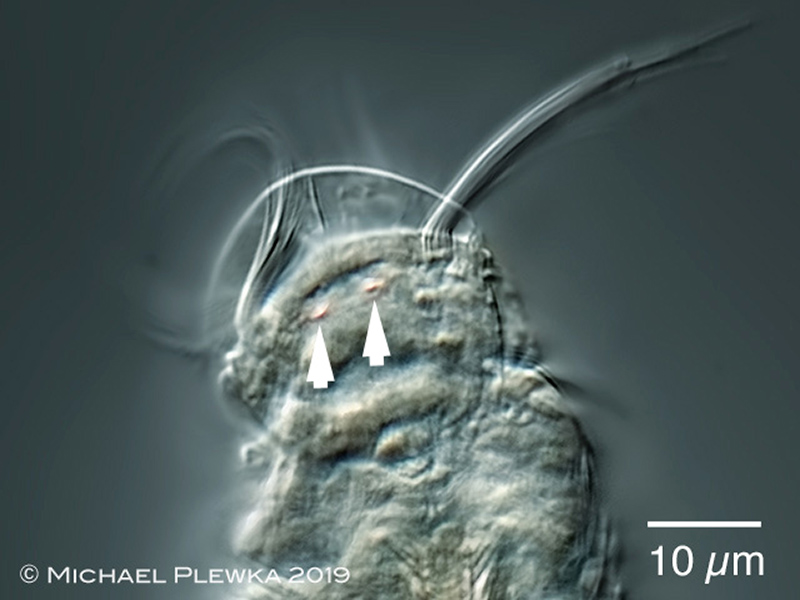
| Bryceella stylata; specimen from (6) showing also 2 light-refracting bodies (arrowheads) |
| |
 |
| Bryceella stylata; crop of above image(6) |
| |
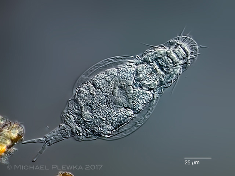 |
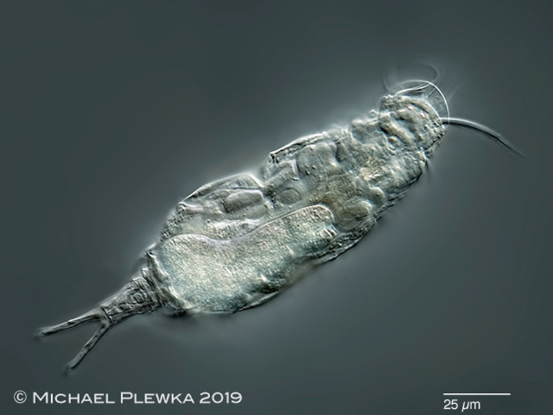
| Bryceella stylata (1); dorsoventral view; focus plane on the rostrum |
| |
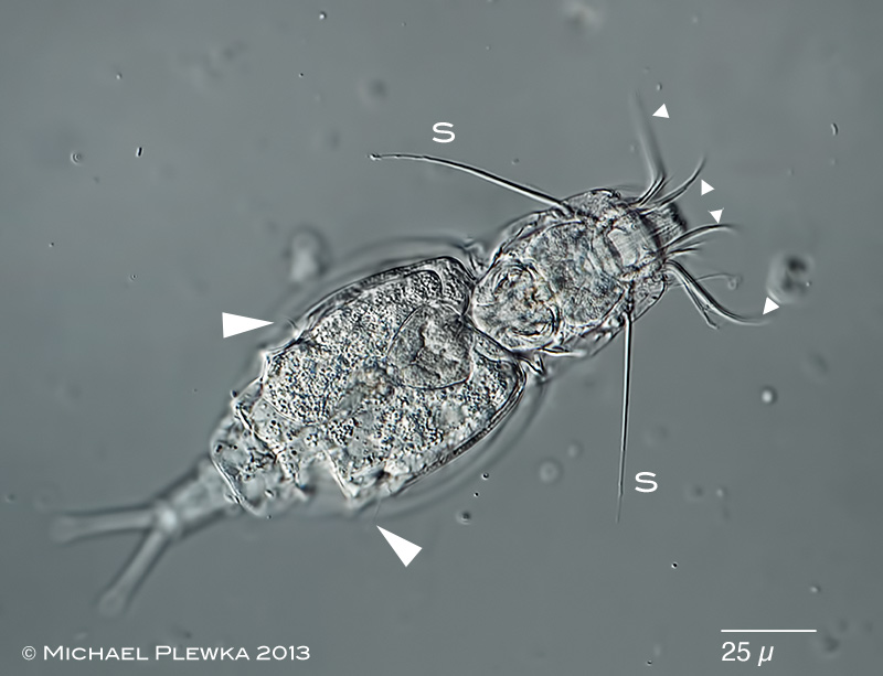 |
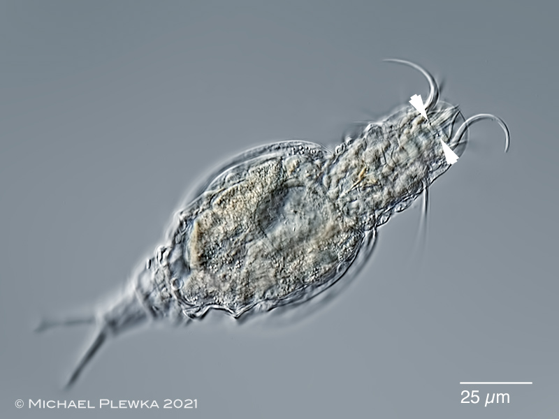
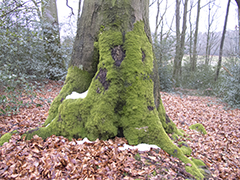
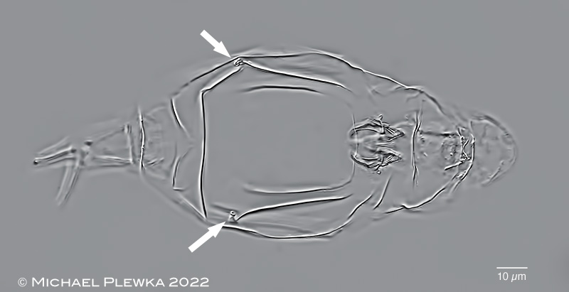
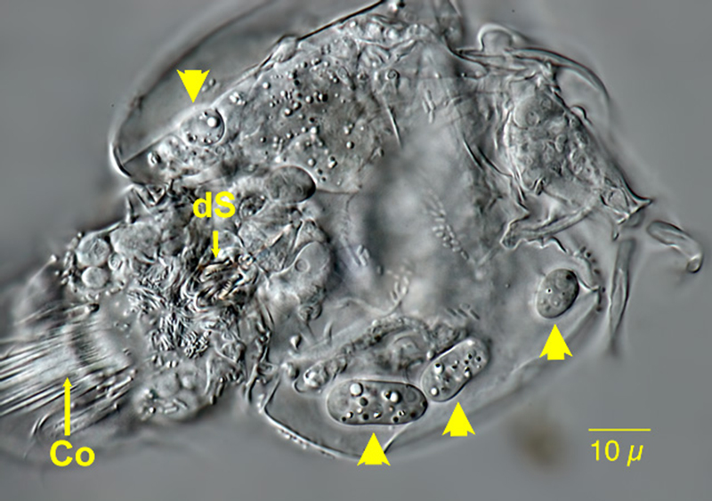
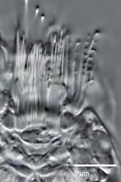
| Bryceella stylata (2); optical median section shows the conspicuous long lateral styli (S) and the apical bristles (triangles). Arrowheads: lateral antennae (Hattingen Oberstueter, tree moss, 13.02.2013). Sympatric with Bryceella perpusilla |
| |
 |
| Bryceella stylata (2); another specimen, vital staining of stomach with neutral red (Hattingen Oberstueter, tree moss, 13.02.2013) |
| |
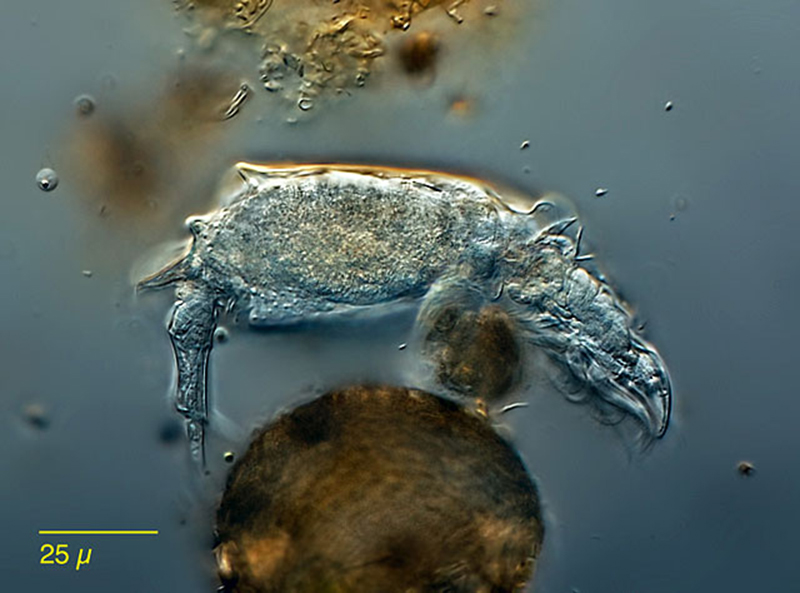 |
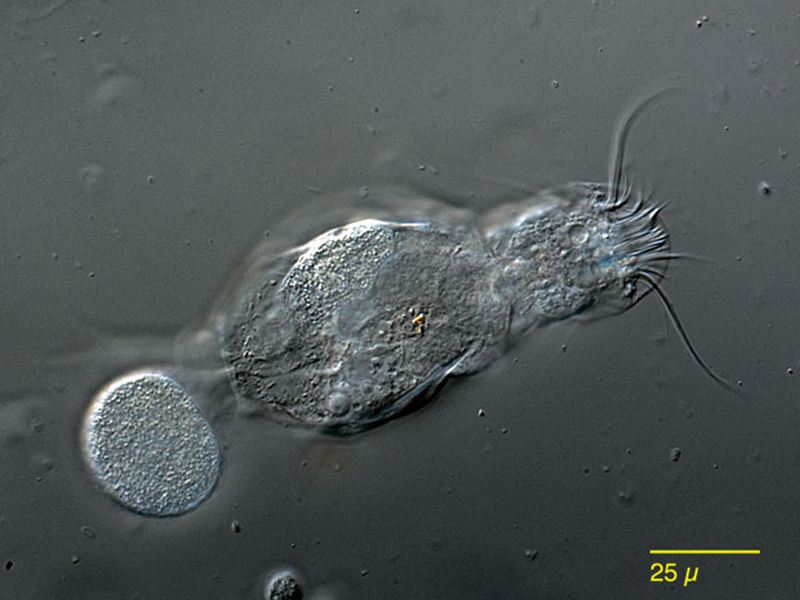
| Bryceella stylata (1); lateral view of a specimen whirling at a small ball of detritus. |
| |
 |
 |
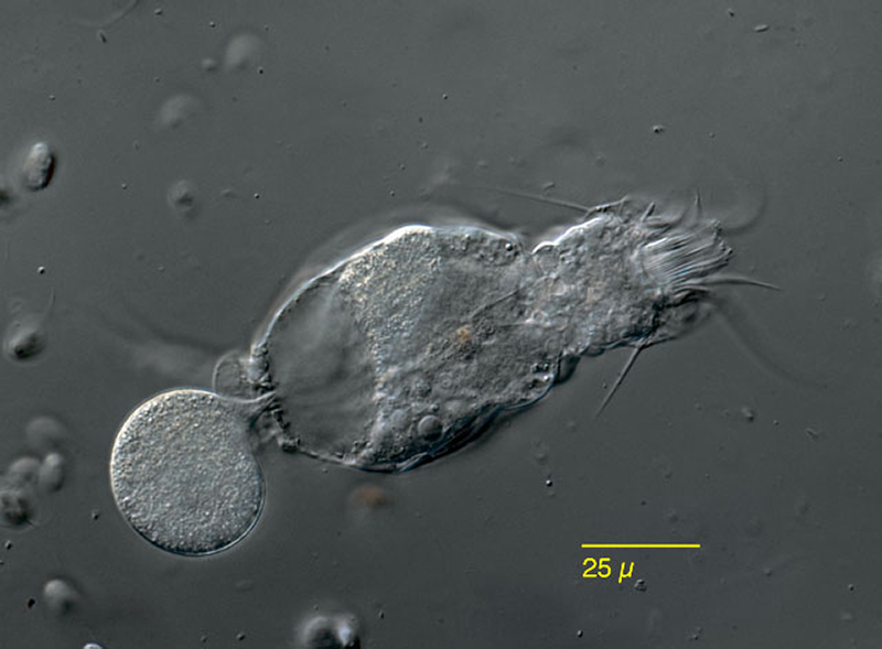
| Bryceella stylata (1); egg deposition; two focus planes. |
| |
 |
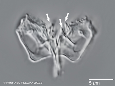
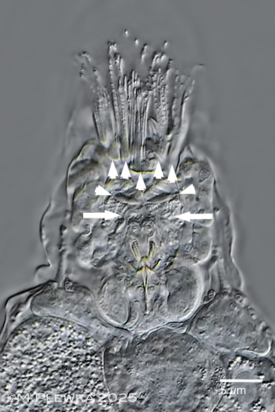
| Bryceella stylata (1); semi-macerated specimen showing the trophi and epipharyngeal structures. The arrows point to the openings of the lateral antennas. |
| |
 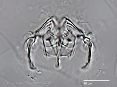 |
| Bryceella stylata (1); virgate trophi >> incus (= fulcrum + rami): rami with teeth on the inner margins (arrows) Left: specimen from 12/2022; right: specimen from Barentsøya 10.10.2017 |
| |
 |
| Bryceella stylata (1); specimens kept in a sphagnum sample for several weeks showed parasites (yellow arrows). (28.12.2010) |
| |
 |
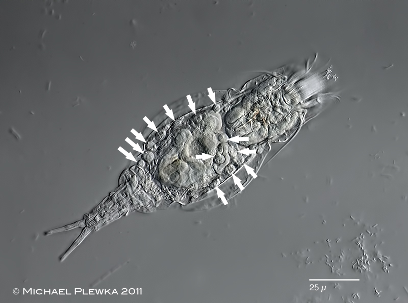
| Bryceella stylata; Detail image showing at least four parasites (Co: cililary organ) (28.12.2010) |
| |
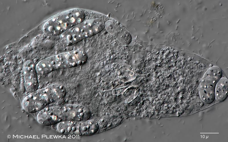 |
| Bryceella stylata; some days later: most of the specimens show infection with parasites. The parasites have conspicuous light refractive bodies. (02.01.2011) |
| |
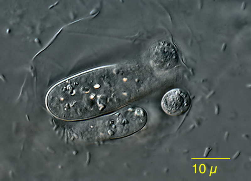 |
| Bryceella stylata; another developmental stage? (02.01.2011) |
| |
 |
 |
| Bryceella stylata; specimen from (5); (lower image: crop of the above image); showing two structures that might be eyespots (arrowheads). |
| |
 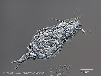 |
| Bryceella stylata; left: lateral view of specimen from (8); right: dorsoventral view of specimen from (3) |
| |
  |
| Bryceella stylata; specimen from (7). Left: the arrows mark the cilia of the buccal field. The cirri of the ventral ciliary field are organised in special areas (?hypodermis cells?; marked by arrowheads.) These cirri show some minute regularly arranged hooks that maybe the bent tips of those cilia that are combined to cirri. Maybe the have a function for locomotion. Right image: detail of some cirri. |
| |
| |
| |
|
|
| |
| |
|
|
| |
| |
|
|
| |
| |
|
|
| |
| |
| |
| |
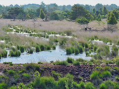
| Location: NSG Heiliges Meer, NRW, Germany, "Seerosenteich" (3); Lanildut, Britanny, France, wall (5); |
| Habitat: between algae (3); dry moss(5); |
| Date: 27.04.2019 (3); coll.: 05.08.2019, img: 07.12.2019 (5); |
|
|